


推荐产品


公司新闻/正文
SPRi 在核酸适配体开发中的应用
人阅读 发布时间:2015-12-02 14:40
(a) Budapest University of Technology and Economics, Department of Inorganic and Analytical Chemistry, Budapest, Hungary
(b) HORIBA Scientific, Palaiseau, France
Aptamers are in vitro-generated, short, single-stranded nucleic acids that selectively bind to target compounds such as small molecules or proteins1, 2. Their main advantage over monoclonal antibodies is their resistance to denaturation and degradation. Being very robust and easy to synthesize, they are becoming widely used for the development of aptamer-based biosensors in the diagnostics field3.
This application note shows that the HORIBA Scientific SPRi platform is suitable for the analysis of aptamer-based molecular interactions. For this purpose, the interaction between human IgE protein, an antibody involved in allergic reactions, and a human IgE specific aptamer4 is presented as proof-of-concept.
Key words:
Aptamer - Aptamer, protein interactions - Human IgE - Kinetics analysis - Affinity - Surface Plasmon Resonance imaging
Material and Method

DNA aptamers
The IgE specific DNA aptamer sequence (5’-GGG GCA CGT TTA TCC GTC CCT CCT AGT GGC GTG CCC C-3’) and the negative control DNA (5’-GGG GCA CGT TTA TTT TTT TTT TTT TGT GGC GTG CCC C-3’) were ordered with TTT TTT T-(CH ) –SH spacer at the 3’ end from Sigma-Aldrich.
Immobilization of the human IgE (hIgE) aptamer on the SPRi-Biochip™
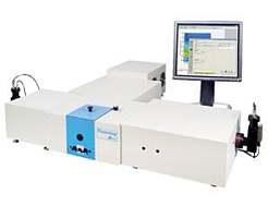
The thiol modified IgE aptamer and reference DNA was immobilized on the planar gold SPRi-Biochip™ sensor through thiol-Au chemisorption.The aptamer solutions were prepared at 10 µM in 20 mM PBS pH 7.4. Four replicate spots were printed simultaneously for each aptamer using the SPRi-CFM (Figure 1) during a 15-minute back-and-forth cycling. The SPRi-CFM uses flow deposition to immobilize 48 molecules in a single run (up to 144 spots per chip). Immobilizing with flow generates increased spot homogeneity and higher immobilization levels. After the immobilization procedure, the SPRi-Biochip™ surface was blocked with a 0.1 mM MUTEG solution prepared in 10 mM PBS pH 7.4. While MUTEG has good resistance against non-specific adsorption, it is sterically short enough not to hinder the specific binding event.
SPRi experiment

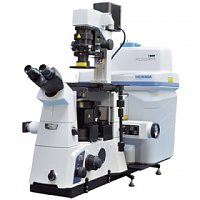
The spotted biochip was inserted into the SPRi-PlexII™ instrument from HORIBA Scientific. The system is equipped with a 200 µL sample loop, a continuous flow pump and an in-line degasser. The flow rate was set to 50 µL/min, and the working temperature was fixed at 25°C. Different concentrations of hIgE prepared in a two-fold dilution in the running buffer (10 mM PBS + 1 mM MgCl ) were injected in order to determine the affinity between hIgE and its aptamer (see Table 1). The SPRi-Biochip™ surface was regenerated using a 12 mM NaOH with 1.2 % EtOH solution after each hIgE injection.
| c0 | c1 | c2 | c3 | c4 | c5 | |
| hlgE concentration (ng/mL) | 0 | 63 | 125 | 250 | 500 | 1000 |
Table 1: Concentrations of hIgE passed over the SPRi-Biochip™ surface
Results and Discussion
Specific aptamer-hIgE interaction
Figure 2 shows a SPRi difference image obtained after the injection of 1000 ng/mL hIgE (concentration c5). Only hIgE aptamer spots become white, which confirms that the aptamer-hIgE interaction is highly specific. The SPRi difference image provides a quick confirmation of the presence of binding and its specificity.
The SPRi signals obtained on the hIgE aptamer spots after the subtraction of the negative control signal were compared for the 5 different concentrations of hIgE (see Table 1). Figure 3 shows the calibration curve obtained; the plot was fitted with a linear model (R² = 0.9842).

Figure 3: Calibration curve obtained for the aptamer – hIgE interaction
Kinetics analysis of the aptamer-hIgE interaction
The SPRi data was then processed using ScrubberGen software in order to determine the kinetic parameters (association k
and dissociation krates) and the affinity (K ) of the aptamer-hIgE interaction. The data was fitted using a 1:1 interaction model (Figure 4). The affinity between the aptamer and hIgE was about 730 pM.
References
1. C. Tuerk and L. Gold, Science, 1990, 249, 505-510.
2. A. D. Ellington and J. W. Szostak, Nature, 1990, 346, 818-822.
3. S. D. Jayasena, Clinical Chemistry, 1999, 45, 1628-1650.
4. T. Wiegand, P. Williams, S. Dreskin, M. Jouvin, J. Kinet and D. Tasset,The Journal of Immunology, 1996, 157, 221-230.
Conclusion
This application note demonstrates that the SPR imaging platform from HORIBA Scientific is well adapted to the characterization of aptamer- protein interactions. The SPRi difference image gives meaningful information regarding the presence of binding and its specificity. The real- time monitoring of the interaction gives access to the kinetics (association and dissociation rates) and the affinity of the interaction. The multiplexing capabilities of the platform will bring very promising applications for the development of aptamer-based biochips in biomedical diagnostics.
Figure 4: Kinetics analysis of the aptamer – hIgE interaction using ScrubberGen

询价列表

暂时没有已询价产品
